- Home
- Products
- CHROME GuidedSMILE
- Patient Records
- Patient Records and Checklists
2. Patient Records
Creating Patient Records
CHROME GuidedSMILE™ patient records are simple but specific.
Incomplete patient records will delay your case.
This page provides all the information needed to successfully capture complete patient records.
For questions about patient record requirements, please call us at (216) 663-2233.
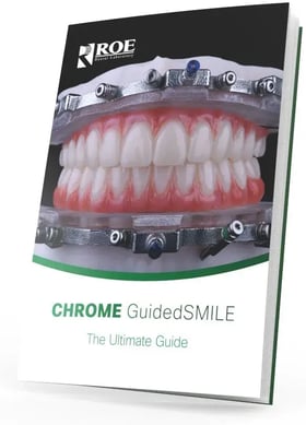
Download Your Free CHROME GuidedSMILE
Ultimate Guide Now
What You'll Gain In This 90-Page Guide:
- Mastery of the 6-component & surgical protocol of the CHROME GuidedSMILE system
- Complete understanding of all necessary patient record requirements on a per-patient scenario
- Comprehensive day-of-surgery prosthesis conversation process
Available in digital or print format (free mailing included).
Single Arch Patient Records
| . | Single Arch Dentate | Single Dentate Arch | Single Edentulous Arch | Single Edentulous Arch |
|---|---|---|---|---|
| Patient Photos | Opposing DENTATE arch | Opposing EDENTULOUS arch | Opposing DENTATE arch | Opposing EDENTULOUS arch |
| Patient Photos | ALL Photos req'd | ALL Photos req'd | ALL Photos req'd | ALL Photos req'd |
| Master Casts / Impressions | Capture ALL land areas of both arches |
• Surgical arch - capture ALL land areas • Non-surgical arch - capture teeth of opposing dentition |
• Capture ALL land areas - non-surgical arch only |
• Capture impression of opposing denture, teeth only |
| Bite Registration | Full occlusion or CR bite | Full occlusion or CR bite | Full occlusion or CR bite | Full occlusion or CR bite |
| CBCT Scans | Scan OPEN position, denture out of the mouth | Scan OPEN position, denture out of the mouth |
• Scan denture in the mouth, in occlusion, not separated • Scan denture alone resting on foam, not plastic |
• Scan denture in the mouth, in occlusion, not separated • Scan denture alone resting on foam, not plastic |
| . | Download Info | Download Info | Download Info | Download Info |
Double Arch Patient Records
| . | Double Dentate Arches | Double Edentulous Arches | Dentate & Edentulous Arches |
|---|---|---|---|
| Patient Photos | ALL Photos req'd | ALL Photos req'd | ALL Photos req'd |
| Master Casts / Impressions | Capture ALL land areas | N/A | Capture ALL land areas - dentate arch only |
| Bite Registration | Full occlusion or CR bite | Full occlusion or CR bite | Full occlusion or CR bite |
| CBCT Scans | Scan BOTH arches, OPEN position, with any removable prostheses out of the mouth |
• Scan BOTH dentures in the mouth, in occlusion, not separated • Scan maxillary denture alone resting on foam, not plastic • Scan mandibular denture alone resting on foam, not plastic |
• Scan BOTH arches with the denture in the mouth, in occlusion, not separated • Scan denture alone resting on foam, not plastic |
| . | Download Info | Download Info | Download Info |
Patient Record Checklists
Click the patient case scenario below to download a ready-to-print PDF checklist of the records necessary.
Single Arch
- Single Dentate arch with opposing Dentate arch
- Single Dentate arch with opposing Edentulous arch
- Single Edentulous arch with opposing Dentate arch
- Single Edentulous arch with opposing Edentulous arch
Double Arch
Single Arch Cases
Dentate arch, opposing Dentate
Dentate arch, opposing Edentulous
Edentulous arch, opposing Dentate
Edentulous arch, opposing Edentulous
Double Arch Cases
Double Dentate Arches
Double Edentulous Arches
Dentate Arch Opposing Edentulous
Simple Patient Records (Webinar Series)
Patient Records Part 1
Patient Records Part 2
Patient Records Part 3
Patient Records Part 4
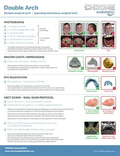
How to Capture the Necessary CHROME GuidedSMILE Patient Records
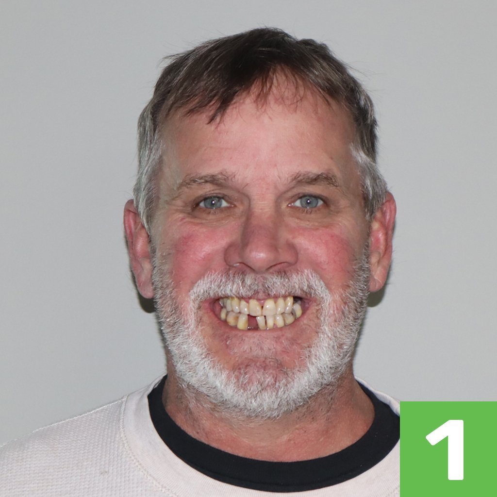
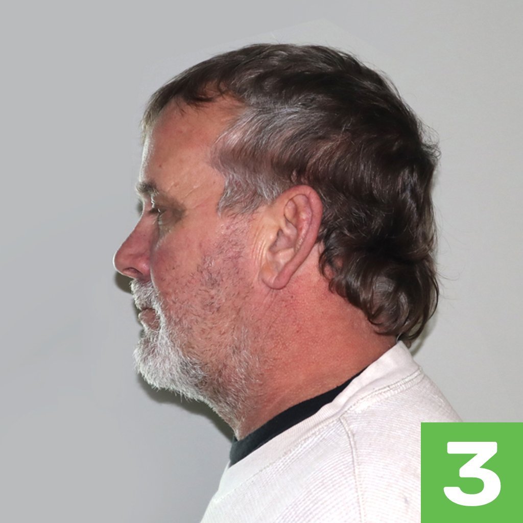
1. CHROME GuidedSMILE Photographs
Good CHROME photos
- Full face full smile
- Full face exaggerated smile
- Full face profile
- Center retracted in occlusion
- Left retracted in occlusion
- Right retracted in occlusion
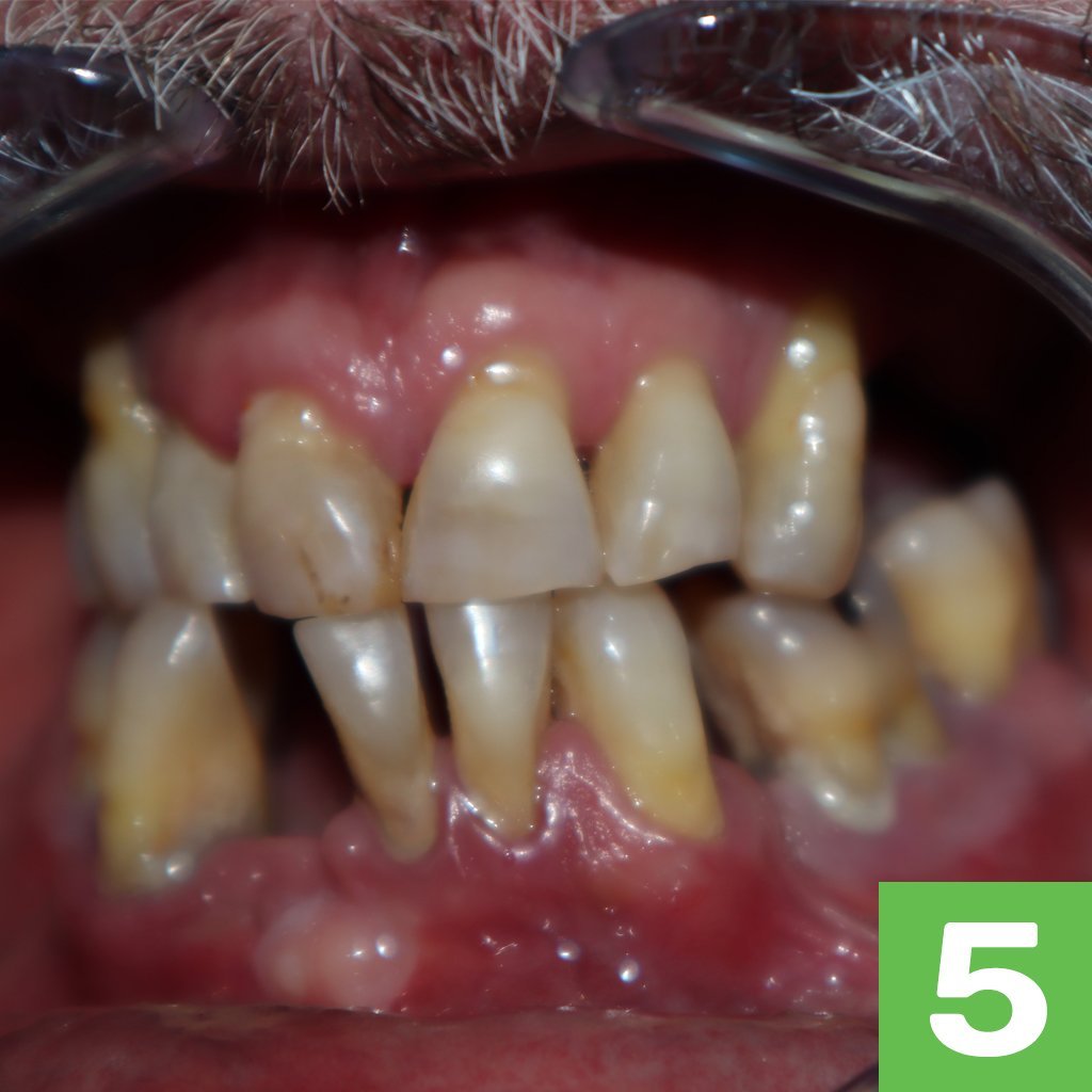

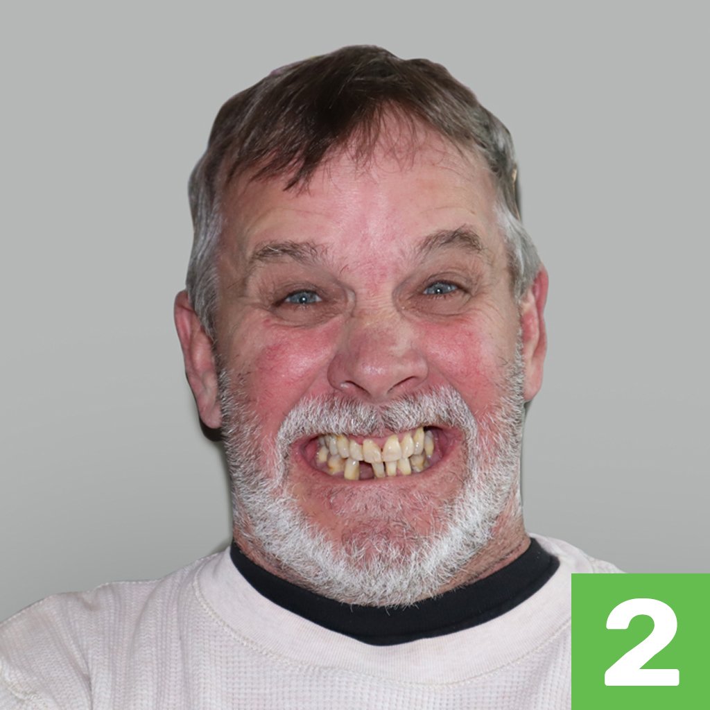
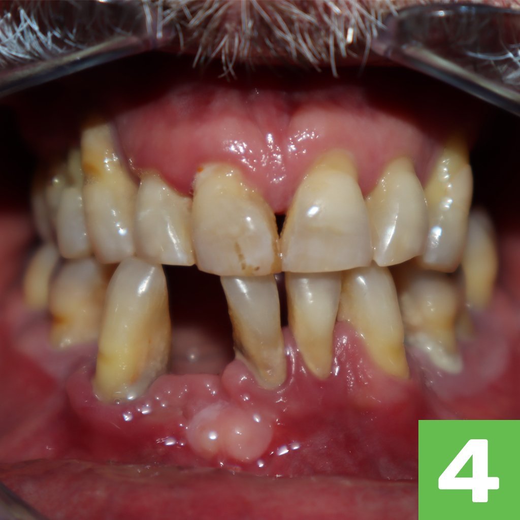
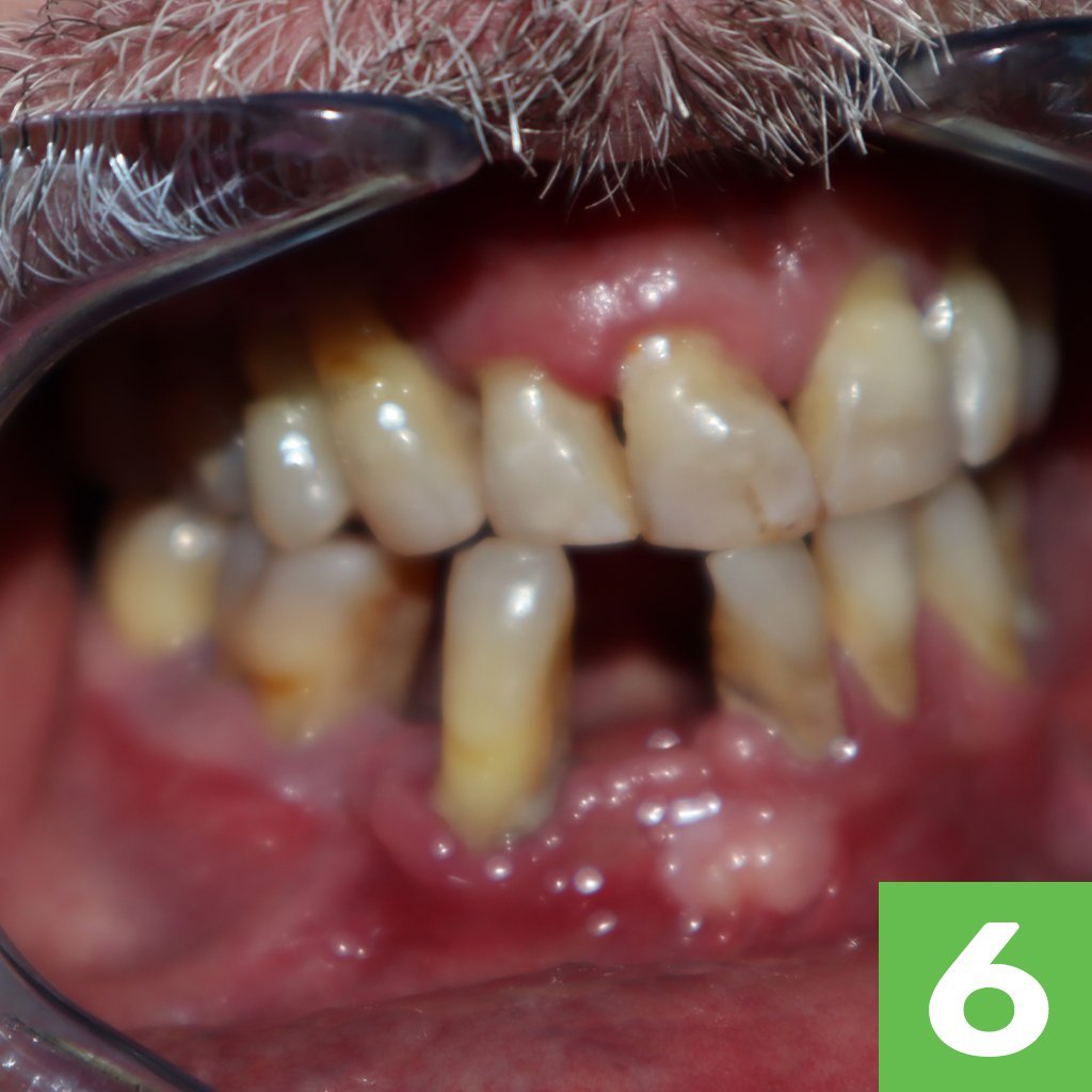
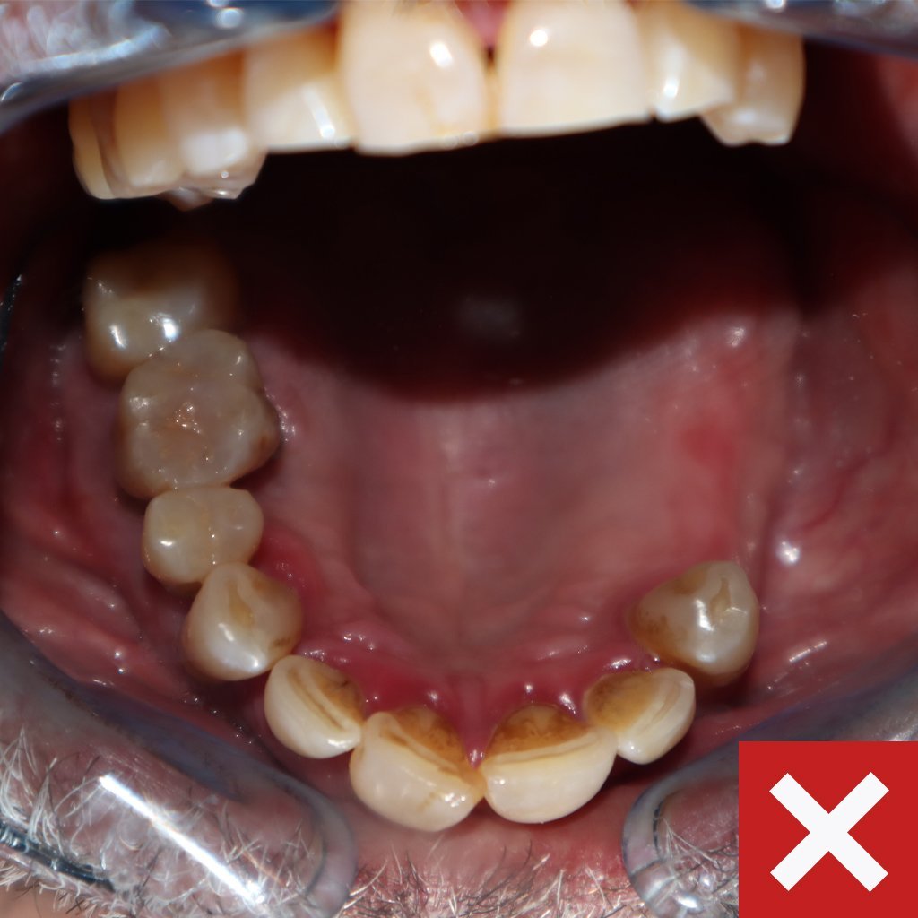
2. Master Casts / Impressions
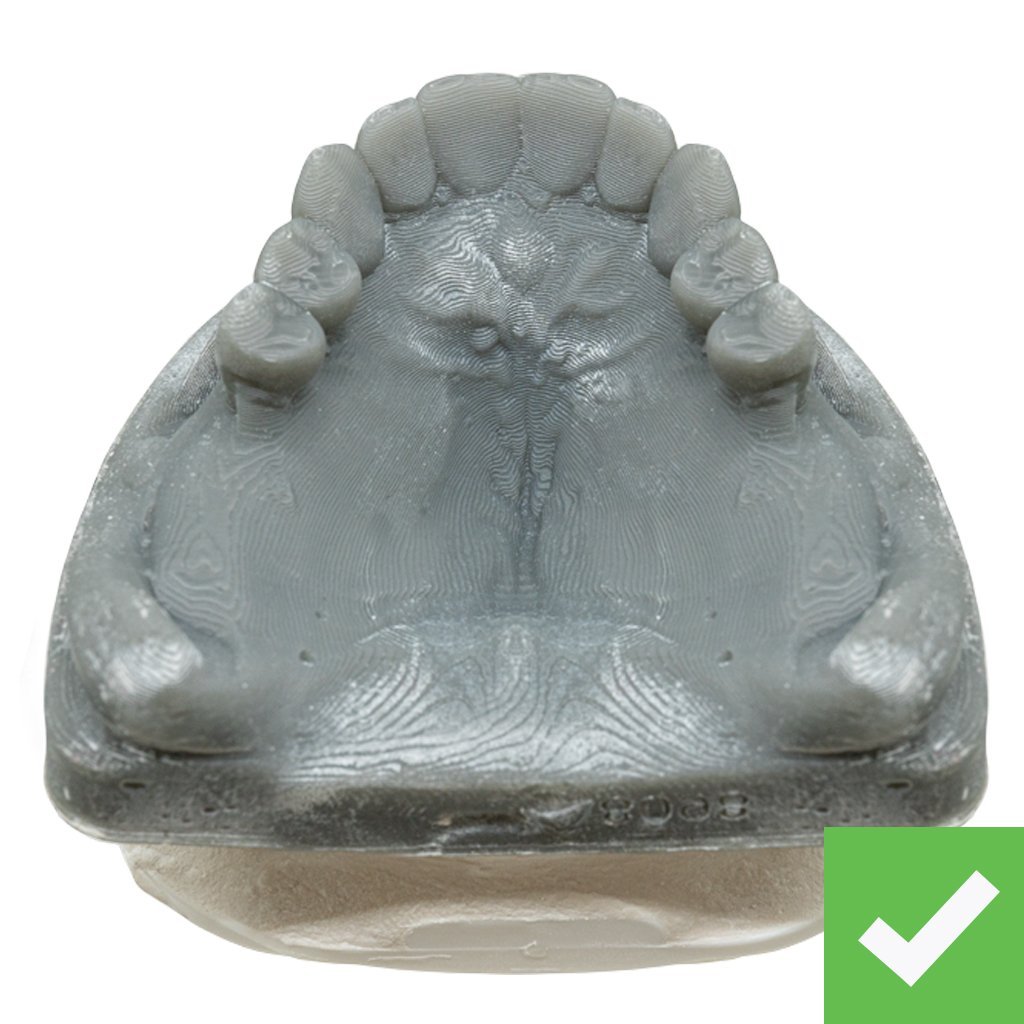
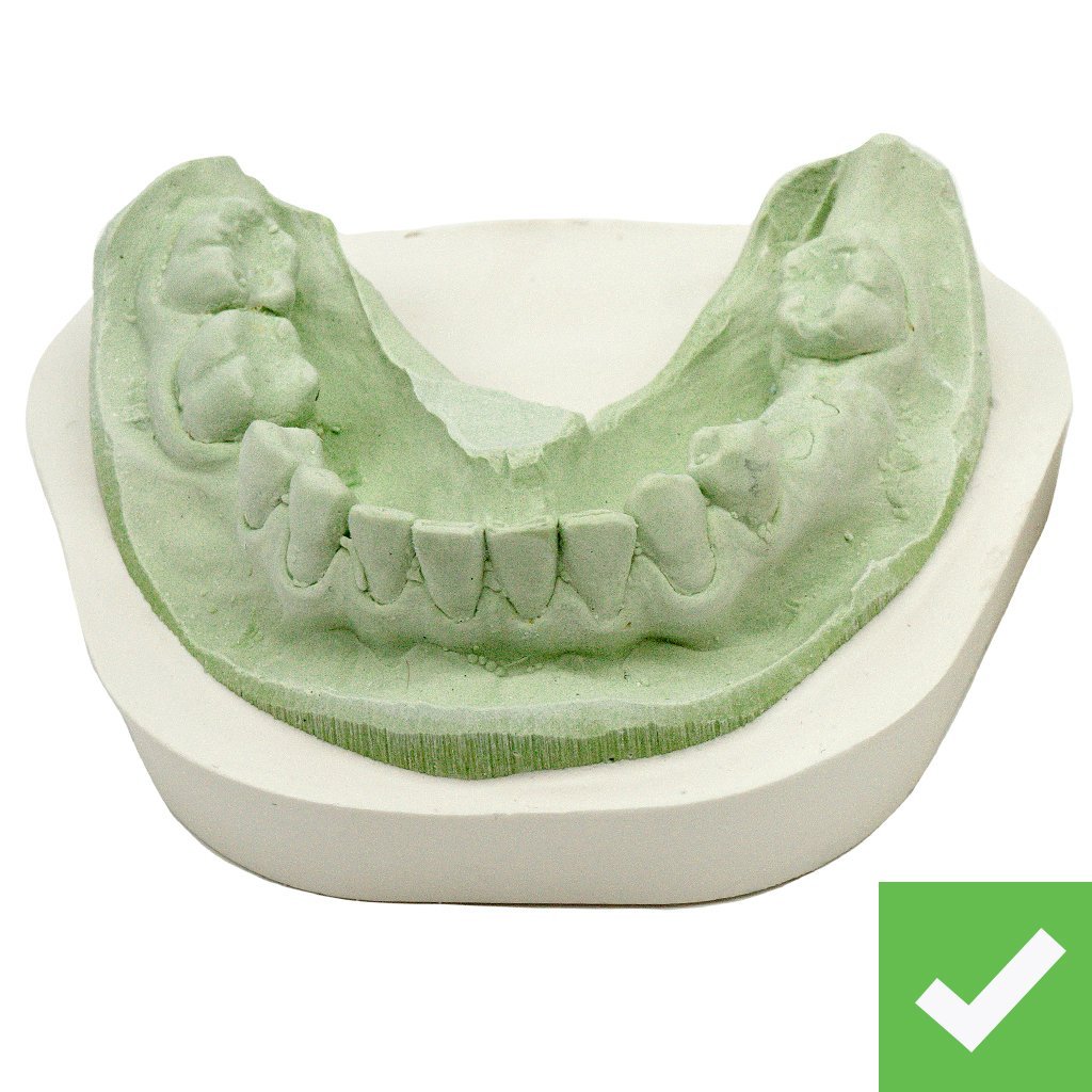
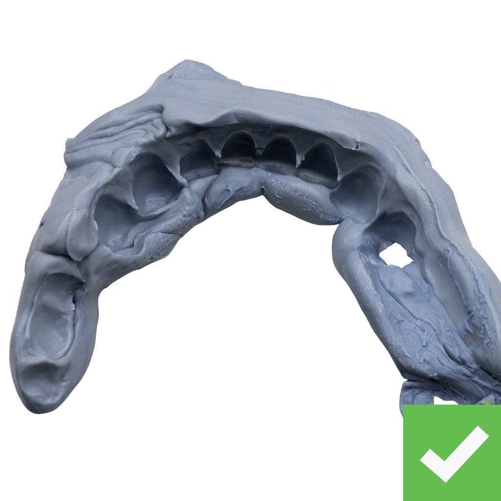
Good master casts/impressions
- Capture full palate
- Capture most/all of the surrounding labial & lingual tissues, the vibrating line, the full roll
- Are free of noticeable holes, skips, voids, or bubbles
Helpful Guidelines
- Most CHROME cases use a backup denture; please capture as much anatomy as you would for an immediate full denture.
- It can be challenging to reach the entire vestibule with an IO scanner, but please try to capture as much of the palate as possible.
- If there is distortion, please retake the impression. It is best to catch inaccuracies at the office.
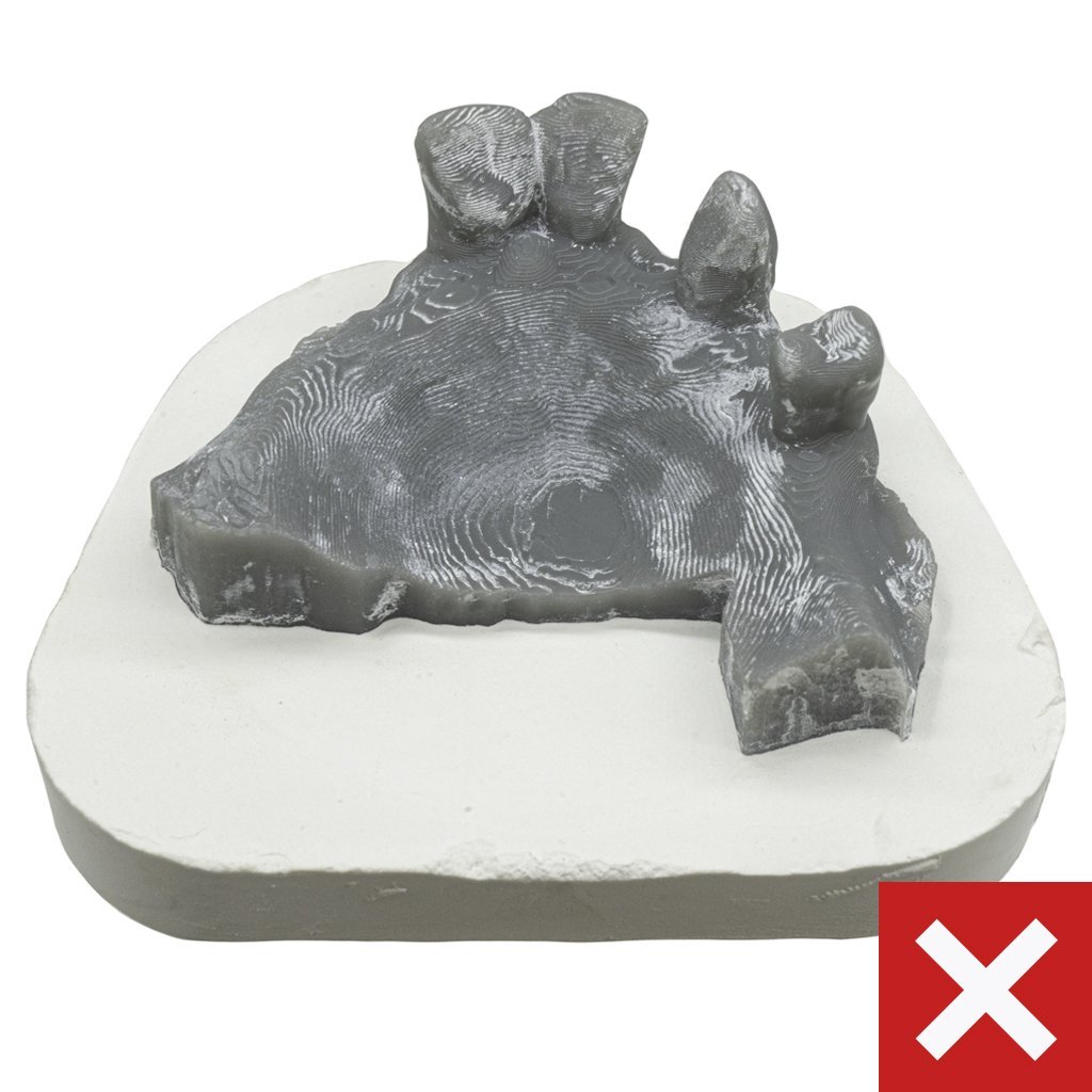
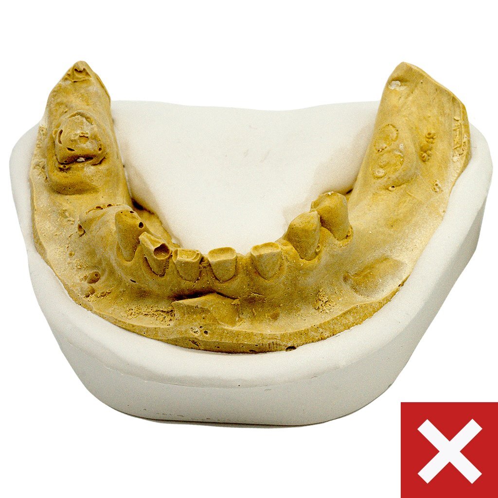
Bad master casts/impressions
We cannot use master casts or impressions that do not meet the desired coverage per patient scenario (see record checklist for details), have distortion, or have noticeable holes, drags, bubbles, and voids.
3. Bite Registration
Good bite registrations
- Capture full occlusion or CR bite.
- Taken with the patient in a closed bite, in natural occlusion.
- Taken with the patient’s prosthesis in mouth, if applicable.
Helpful Guidelines
- Use enough material to capture the occlusion, but not too much as to open the bite.
- Neuromuscular bites are accepted as well; these are often the best, most accurate bites.
- If the patient is missing posterior stops, ROE recommends bite blocks or a JC Try-In. A JC Try-In appliance will stabilize the bite and help establish occlusion.
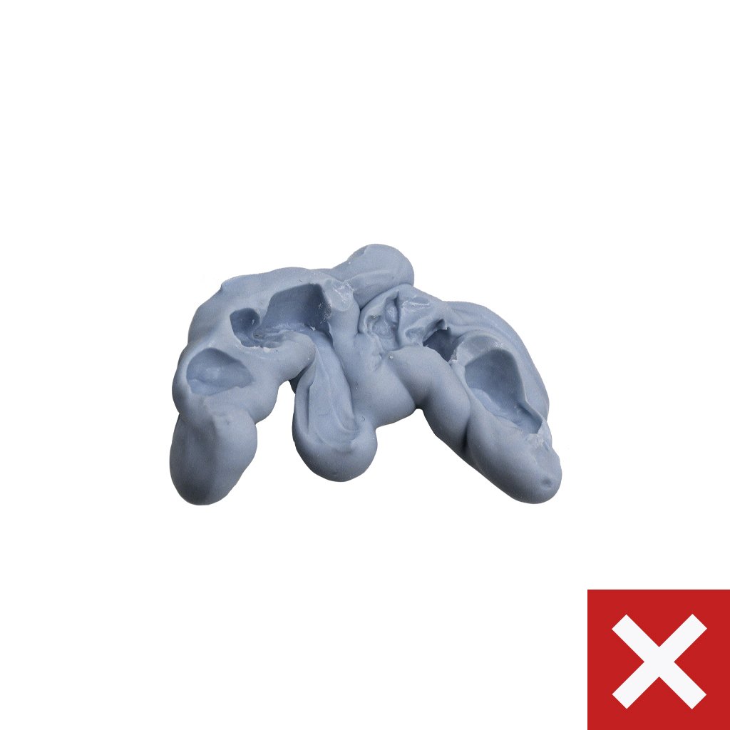
Bad bite registrations
We cannot use bite registrations that:
- Only capture a narrow centric occlusion.
- Used too much material, which separated the bite.
- Were not captured with teeth in natural occlusion.
Scanning Videos
Dual Scan Technique
Scan a Denture and Bite
Scan a Relined Denture
4. CBCT Scans
Dentate
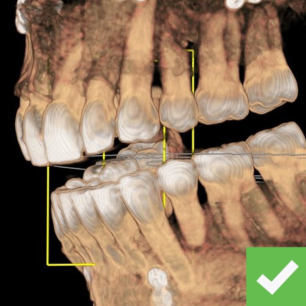
Good dentate CBCT scans
- Capture both arches.
- Taken with the patient in an OPEN position.
- Taken with the patient’s denture(s) out of the mouth, if applicable.
Helpful Guidelines
- Have the patient bite on cotton rolls or the bite fork.
- Place cotton rolls between cheeks and teeth for a better image.
- If the field of view only allows one arch, send two individual scans of each arch.
- Export DICOM files (.dcm) from the CBCT into a patient folder- this should contain 200-400 individual .dcm files. Zip the patient folder and upload with Rx.
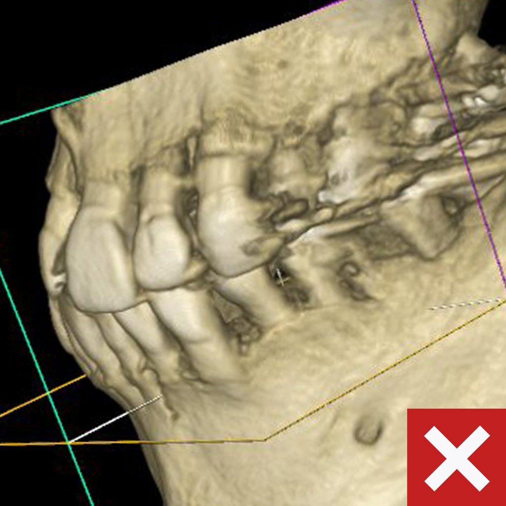
Bad dentate CBCT scans:
We cannot use dentate CBCT scans that:
- Captured the patient in a closed position.
- Captured the patient with their denture(s) in the mouth.
Edentulous
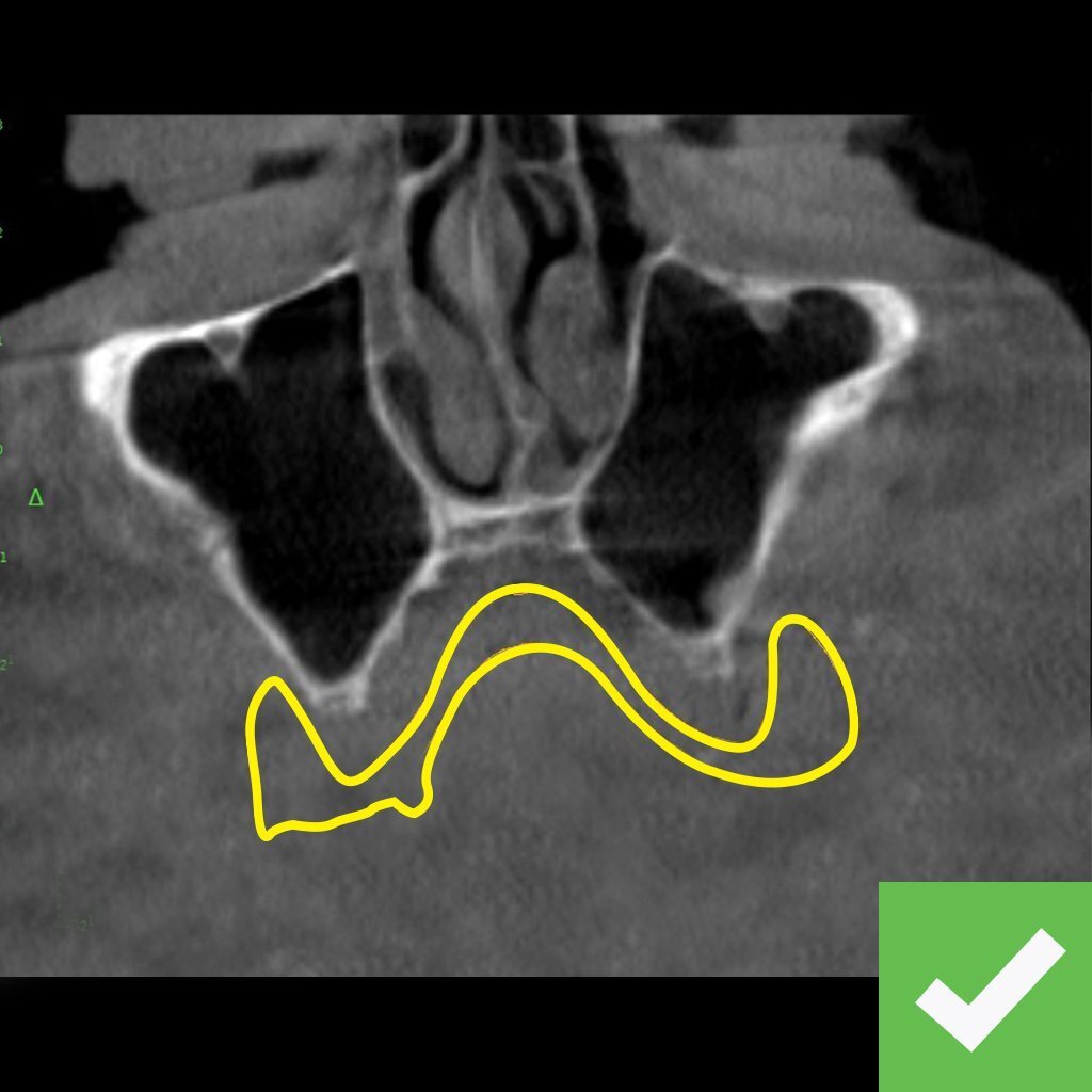
Good dentate CBCT scans
- Capture both arches.
- Taken with the patient in an OPEN position.
- Taken with the patient’s denture(s) out of the mouth, if applicable.
Helpful Guidelines
- Have the patient bite on cotton rolls or the bite fork.
- Place cotton rolls between cheeks and teeth for a better image.
- If the field of view only allows one arch, send two individual scans of each arch.
- Export DICOM files (.dcm) from the CBCT into a patient folder- this should contain 200-400 individual .dcm files. Zip the patient folder and upload with Rx.
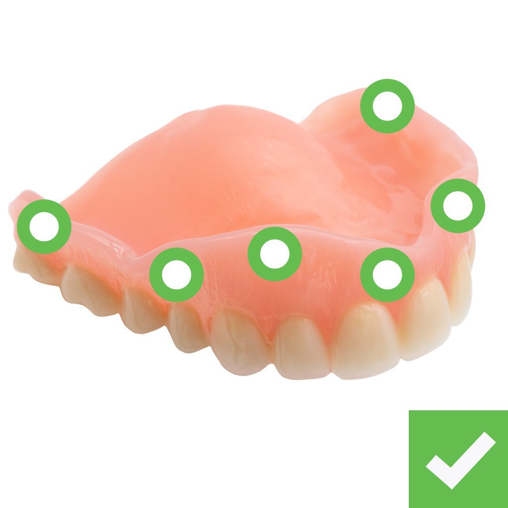
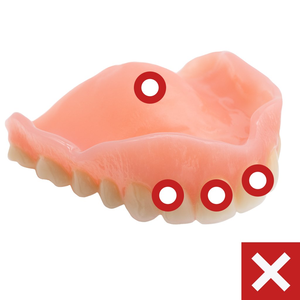
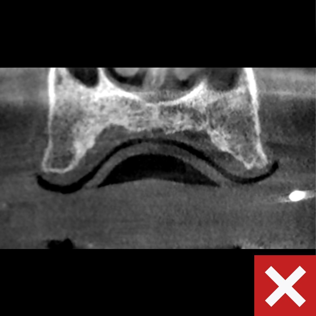
Bad dentate CBCT scans:
We cannot use dentate CBCT scans that:
- Captured the patient in a closed position.
- Captured the patient with their denture(s) in the mouth.
How can we help you today?
Can't find what you're looking for? No problem. Let us help.
ROE’s experts are dedicated to elevating your dental practice.
- Full-service national dental laboratory
- Comprehensive Certified Dental Laboratory (CDL) services
- Case planning support from 20+ Certified Dental Technicians
- Specialists in both fixed, removable, implant and full-arch dentistry
- Tailored support for all cases, from routine to complex
- National Association of Dental Laboratories (NADL) member
- Only FDA-approved materials used
- All products made in the USA
- Thousands of free online education materials

